JJIREVC

Ultrasound Guided FNAC is an advanced diagnostic procedure where Fine Needle Aspiration Cytology is performed with real-time ultrasound imaging. This technique allows precise needle placement into the target lesion, especially when the lump is small, deep, or not easily palpable. Ultrasound Guided FNAC improves diagnostic accuracy by ensuring that the correct area is sampled.

Accuracy is critical in diagnostic testing, and Ultrasound Guided FNAC plays a key role in achieving reliable results. It is commonly used for thyroid nodules, lymph nodes, breast lesions, and deep-seated masses. By visualizing the lesion during the procedure, Ultrasound Guided FNAC reduces sampling errors and the need for repeat FNAC procedures, leading to better clinical outcomes

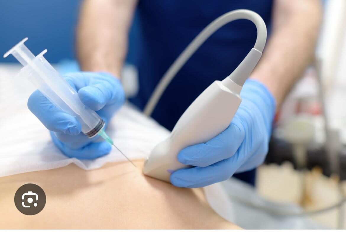
During Ultrasound Guided FNAC, ultrasound imaging is used to locate the lesion clearly. A fine needle is then guided into the target area under continuous visualization. Once the needle is positioned accurately, cells are aspirated and sent for cytological examination. The procedure is quick, safe, and causes minimal discomfort, with no need for anesthesia or hospital admission.

Ultrasound Guided FNAC offers high diagnostic accuracy with a low risk of complications. It helps clinicians differentiate benign and malignant conditions with confidence. Because the procedure is minimally invasive and image-guided, patients experience minimal pain and can return to normal activities immediately. Ultrasound Guided FNAC is a trusted diagnostic tool in modern pathology practice.

Leave a Reply

Your email address will not be published. Required fields are marked *